Royal Raymond Rife And His Amazing Microscope
 By Gerry Vassilatos
The Microscope. The Super Microscope. There were predecessors to the prismatic marvel of Dr. Royal Raymond Rife, but no equals. Others had designed and used oil immersion lenses, dark-field illuminations, and deep ultraviolet light, each holding part of the secret for optically magnifying infinitesimal objects. But the design which Dr. Rife developed outpaced all of these.
It is doubtful whether you have ever heard his name. Reasons for mass forgetfulness run deep. Truths have been kept from you. Only a careful and relentless study of the past will relinquish secrets purposefully and cunningly buried. The information is safely nestled in dust laden libraries which few now venture to search. Perhaps you will recognize why his name has been blotted out of the historical records before we reach the end of this amazing biography.
Dr. Rife began as a research pathologist. A medical crusader of the very highest qualifications, is was a heart filled with but one goal: the eradication of disease. Dr. Rife recognized first and foremost that successful medicine relies on vision, on light. What we cannot see we cannot battle. An unseen foe is impossible to destroy. Therefore his first quest was to secure a vastly improved system of microscopy. Once he could see, once all could see, then the pursuit of medical knowledge could again move forward. An armada of equipped seers could assail the foe on every shore. Looking for more light.
Dr. Rife’s study of microscopy detailed every component and premise which tradition had presented to our century. The creation of a super microscope would run counter to every physical law and restriction which the previous two centuries had accumulated. Academes began again to love the writing of papers. Without the exercise of experiment, however, all these papers were so much tinder.
Dr. Rife wanted to develop super microscopes capable of seeing viruses. His aim was to chart and catalogue them, understanding that these represented a deadly foe which exceeded bacilli in their destructive assault on humanity. His quest now began. He reduced the fundamental premise by which microscope design had developed, analyzing each separate component and premise.
FOCUS
Optical designers had been adding ever more complex components to the design which began with van Leeuwenhoek. Lenses were compounded to lenses, crowns were added to compounds, crowns were added to crowns …the complexity was frightful. Simplifying the problem necessarily led to Rife back to the study of optical geometry and the comprehension of simple ray divergence.
Rife thought on these ancient principles. An ideal magnifying system is a geometric construction of extreme simplicity. Diverging light rays can magnify any object to any magnification. Given a strongly divergent light source and a great enough distance, one can theoretically magnify the indivisible! This is the principle which underlies projection microscopy. Dr. Rife realized that the projection microscope represented the best and simplest means for magnifying infinitesimal objects. One simply needed to discover a means by which a vanishingly small, brilliant radiant point could project divergent rays to the surface of any material speck. No virus, however indistinct and cunning, could hide from such an optical magnifier.
The theoretical design of microscopes relies purely on geometric principles. Actual materialization of these principles requires material manipulation, since geometric rays and light rays are significantly distinct. What is a microscope essentially? What is achieved in a microscope with light rays? The notion is quite simple. Take divergent rays from a vanishingly small point of brilliant emanations and allow them to pass through any specimen which is to be viewed. Light from this encounter is then made to diverge as far apart as possible in a given space. Geometrically it is possible to divert rays from a vanishingly small point out to an infinite distance. This geometric construction would produce unlimited (ideal) magnifications. Provisions toward this ideal goal would require that the point radiant source be tiny and brilliant enough, the specimen be close enough to the radiant point, and the image diverging space be very long. The geometric divergence of the point light source is the magnification factor. But geometry is an idealized reality. And ideal geometry encounters significant frustrations when implementing light in inertial space.
The most basic type of microscope is the projection microscope. It is the most simple system which is employed to greatly magnify the most infinitesimal objects. In the more common version, light is made to pass through a tiny specimen. Light from the specimen is forced to diverge across a long space by means of a very small focussing lens. Rays from this lens cross, diverging and expanding across a long space. This widely divergent beam is then projected onto frosted glass. The viewing of images derived through these means is indirect, but provides superior magnifications with ultrahigh resolutions.
Formerly, laboratories required compact units capable of close personal manipulations. The development of fine optical microscopes became frightfully complex when more powerful but compact models were required. The notion of a compound microscope is to physically compress the long projection space into a compact tube, delivering the shrunken design to customers who wish to conserve space. The “problem” with compact optical microscopes was bending the necessary wide beam through a small space. The “trick” in a compound microscope was to keep the image rays from prematurely diverging between lens stages.
The long expansion space required for divergent beam magnification had to be “folded” and “convoluted” within imaging tubes. Large numbers of lenses performed this duty. Being thus “convoluted” by lenses to achieve magnification, images produced by most expensive bench microscopes were inherently limited. Since the diverging image in these microscopes is “interrupted” within a greatly shortened space by means of several optical stages, it cannot produce great magnifications with either clarity or brilliance.
Each optical stage continually bends the image until a tremendous effective divergence is achieved. The effects are dramatic, but the necessary stages introduce optical resistances by which magnifications are inherently limited. Fundamental problems with white light alone complicated the problems which designers faced. Breaking into spectral components, each color refused to focus in exactly the same point. As a result, chromatic aberration blurred every image.
The light-crossing action of each lens brought widely diverging light beams into the ocular lens. It was pivotal that these rays be parallel. Images lost most of their radiant power against the tube walls before arriving in the final ocular. Therefore more corrective lenses were added in the beam path to bend the light back from the tube walls. Differences when light travelled between lenses and air introduced more aberrations. Batteries of corrective lenses, crowns and compounds, so loaded the light path with crystal that images lost their original brightness. These horrendous optical problems were never completely solved, despite the high cost of these instruments.
All of these optical horrors were the result of an old tradition which yet compels designers to maintain familiar outward forms. The projection microscope is so simple and potent, one wonders why newer designs had not been developed with as much dedication and zeal. It was the outward form which compelled the convolution of projection microscope simplicity, detracting from the excellence of magnified images. What was really lacking in optical microscopy was the development of true, tiny radiant points of monochromatic light. These diverging ray sources could produce novel and economical projection microscopes.
The numerous optical components of most excellent laboratory microscopes are configured to prevent image splitting, image incoherence, and other optical aberrations. All the differences between geometric ideal are suddenly and severely limited when using light and glass. The optical ideals come short of the geometric ideal.
Geometric rays do not fade with infinite distance. Light rays do. Geometric rays do not blur at their edges with increasing divergence, Light rays do. Geometrically magnified lines do not diminish in their intensity. Light images do. A successful optical approximation to the geometric ideal would produce a super microscope. Dr. Rife decided to manipulate all the possible variables in order to approach, as closely as possible, each part of the ideal geometric construction. If such a feat could be accomplished, he would have successfully bridged the gap between optical and electron microscopy.
POINTS
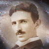
To be sure, numerous individuals had accidentally discovered enormous magnification effects while experimenting in completely different fields of study. A magnifying system which magnified much smaller infinitesimals than viruses appeared in 1891. Nikola Tesla developed a remarkable carborundum point vacuum lamp and made an accidental observation which opened a new world of vision to science.
Tesla began inventing single wire vacuum lamps for purposes of illumination. These were large glass globes powered by very rapidly impulsed currents. The impulse currents made the single supported wires glow to white brilliance, melting them. Impractical for public use, he sought to alleviate this condition by using special crystals. High melting points were required. An assortment of such materials were poised at the single wire termination. When electrified, they suddenly became radiant.
His experiments included using diamond, ruby, zircon, zirconia, carbon, and carborundum. He found it possible to blast the natural gems after a few seconds’ electrification time. But before exploding, each of these crystalline terminations released puzzling patterns of light across the globe surface. This symmetrical pattern of points attracted Tesla’s attention. They appeared when the current was turned on for just an instant.
Moreover, Tesla noticed that the brilliant fixed points of light remained in fixed positions each time he applied the current. Equally astounding was the fact that each material portrayed distinctive point symmetries upon the glass enclosure. The most resilient and successful crystalline material was carborundum, which he ultimately adopted for practical use. This too gave its characteristic point symmetry across the globe.
Tesla was not sure what he had discovered. He intuitively surmised that these point patterns of light somehow revealed the crystalline structure of the excited material. He also utilized the geometrical construction to obtain his deduction. His thoughts turned to the internal crystal conditions. As electrically charged particles were propelled and ejected through the carborundum, they were deflected by infinitesimal points. Diverging from such infinitesimal points, they impinged upon the inside spherical globe which housed the carborundum point. These brilliant points of light were always of the same symmetry because the ejected particles were passing through a fixed grating: a crystalline grating.
He theorized that this fixed pattern represented the greatly magnified crystalline symmetry. This simple apparatus was the world’s first point-electron microscope. The phenomenon responsible for the defined projection of crystalline spaces is referred to as “field emission”. Later, others would duplicate these same results with other crystalline specks. The remarkable X-Ray photography of Max von Laue already permitted the sighting of crystalline atoms. In this scheme a thin crystal point was placed at a critical distance from an X-Ray source. Entering and passing through the crystal slice, divergent X-Rays produced a greatly magnified image of crystal atoms on photographic negatives.
The result of von Laue’s experiment was astounding, but was a purely geometric consequence. Divergent rays from a vanishingly small radiant point can theoretically magnify equally small specks to immense size. But while both Tesla and von Laue produced wonderful results with particle-like emissions, the practical achievement of these ideals were diminished when using optical light rays.
Emile Demoyens (1911) claimed to have seen extremely tiny mobile specks under a powerful optical microscope …but only at noon during the months of May, June, and July! Colleagues thought him quite mad, but Dr. Gaston Naessens has comprehended why these specific time periods permitted such extreme viewing. During these seasonal times the noonday sunlight contains great amounts of deep ultraviolet light. The shortened wavelengths provide a sudden optical boost, permitting the observation of specks which are normally invisible.
Progress in optical science seemed limitless and free. It was anticipated that no limit could bar humanity from viewing the very smallest constituents of matter. But when the physicist Ernst Abbe challenged the high hopes of optical science by imposing certain theoretical limits on optical resolution, all these hopes seemed to dissolve. Abbe claimed that optical resolution depended entirely upon incident light wavelengths, the limit being one-third of the light wavelength used to illuminate the specimen. According to Abbe, the extreme ultraviolet light of 0.4 microns wavelength could not be used to resolve the details of objects smaller than .15 microns.
This theoretical “death-knell” discouraged most optical designers of the time. Since, he claimed, resolution of optical microscopes was restricted from 1600 to 2500 diameters, developing newer optical microscopes was a futile pursuit. Since resolution is the ability of a magnifying instrument to identify details and ultrafine levels of internal structure, the Abbe limit imposed a serious halt on the development of newer optical microscopes.
Continual medical progress rides entirely on the excellence of its instrumentalities. In the absence of new and excellent optical instruments of greater precision, medical progress grinds to a screaming halt. When this happens, academes write papers in the absence of true vision. True knowledge, reliant on vision and experiment, is replaced by unfounded speculation.
Others conceived of electron microscope designs, taking advantage of the Abbe restriction for lucrative purposes. These developers were not good planners, failing to recognize that electron microscopy would place equally grave limitations on biological researchers. Electron beams kill living matter. Magnifying images only after killing them, no living thing could ever be observed in natural stages of activity through electron microscopy. But, if money was to be made, then “all was possible”. Despite the protests of qualified medical personnel, RCA continued its development with Zworykin at their helm.
Electron microscopy, rationally impelled by the Abbe limit, became the new quest of young financiers. Despite the protests of major researchers, RCA continued its propaganda campaigns. This technological imposition, were it developed into a marketable product line, would severely handicap the work of every medical researcher. Pathologists would be literally forced to accept the limitations of the anticipated electron microscope.
Bracing themselves for the announcement of mass-produced electron microscopes, corporate researchers prepared themselves for the laboratory adaptations they would be forced to adopt. Manuals were already being distributed. They would be unable to watch progressive activities in the boasted “highest magnifications ever achieved”. Before RCA reached the goal however, others had already challenged the capabilities of electron microscopy. The unexpected development temporarily threw RCA off balance. The competitors had challenged the Abbe limit, and seemed to be optically working their way into realms in which RCA had claimed “exclusive” rights.
DEEP VIOLET
Vibrating above the deep ultraviolet range were the X-Rays of von Laue’s projection microscope. But this realm was not good for pathologists since XRays would only reveal the structure of crystalline substances. Some designers went ahead and built soft X-Ray microscopes. These devices placed heavy requirements on the preparation of specimens. X-Rays passed right through specimens and would kill them if they were alive to begin with. The very best X-Ray images of tiny specimens required organism-killing metallic stains. Biologists needed to see their specimens in the living state.
While engineers at RCA were yet scrambling to take the competitive edge and seize the new market, several designers of ultra-microscopes began to successfully challenge the Abbe limit. Abbe stated that the maximum resolving power of any ultraviolet ray microscope would be restricted from 2500 to 5000 diameters and no further. But ultra-microscopes constructed by Graton and Dane (Harvard University) succeeded in developing resolutions of 6000 diameters with magnifications of 50,000 diameters.
Dr. Francis Lucas of Bell Telephone Labs developed a modified version of this system in which a maximum magnification of 60,000 diameters was developed. Not only did this work significantly reduce the theoretical limits set by Abbe, but the ultra-microscope which Dr. Lucas designed actually empowered Bell Labs to compete with RCA in the microscope field. Dr. Rife had previously achieved resolutions of 6000 diameters with resolutions of 50,000 diameters. And now, Dr. Rife believed he had a means by which these preliminary feats could be greatly outperformed. The Abbe Limit, a theoretically perfect expression, was dissolving before the new empirical evidence.
Of course, RCA ultimately outdid the propaganda campaign for their own electron microscope system, wiping out the optical systems of both Bell Labs and Harvard University. Nevertheless, independent researchers preferred these ultraviolet microscopes to any system which RCA could market. Attractive because the ultraviolet microscopes permitted life-active observations, pathologists were not impressed by the extra magnifications of electron microscopy.
OBJECTIVE
Ultraviolet light for ultra-microscopes is an absolute necessity. The successful operation of any such device depends on deep UV rays. Monochromatic ultraviolet sources prevented many of the familiar optical aberrations common to optical microscopy. Blurring and fringe degeneration when passing through the optical resistance of lenses would be minimized. The ultraviolet source would also need to be of the shortest possible wavelength in order to approach the geometric ray ideal.
All optical components in the ultra-microscope would then have to be composed of pure quartz crystal in order to flawlessly transduce the deep ultraviolet rays. Even the specimen slides were made of thin quartz glass. The ultra-microscopes of Dane, Graton, and Lucas used as few lenses as possible, being virtually pure projection microscopes.
According to Dr. Lucas, resolution one-tenth of the illuminating light wavelength was obtained. This broke the so-called Abbe optical restrictions by an order of 300 percent; the resolution being brought up to .05 microns. How was this possible? Drs. Dane and Graton further stated that far greater resolution could be obtained through lenses than claimed by their manufacturers. The reason for this? So long as the manufacturers had accepted the theoretical limits there was no incentive toward progress in the field. No one bothered to find out!
The ultra-microscopes demonstrated beyond question that lenses do in fact surpass theoretical limits. The manufacturers, eager to maintain credibility in the academies, had simply endorsed whatever the physicists wrote. Equally as significant was the fact that each of these ultra-microscopes did not require the fixation of specimens before viewing. The embodiment of each ultra-microscope gave new drive to researchers who wished to see live pathological stages in tissue cultures. The systems were immediately demanded and obtained by numerous serious research institutes on both sides of the Atlantic.
Certain highly respected researchers came to believe that the most basic laws concerning physical light were fundamentally flawed. Perhaps light was of an entirely different nature than supposed. This, they mentioned, was why the Abbe limit was such a distorted mathematical expression. Light was not what the physicists declared it to be. This is why Abbe’s assessment was so obviously flawed. But what other assumed truths were holding back fresh discovery? Empirical observations now replaced the theoretical piles with discoveries which were once termed “unlikely” by qualified authorities.
When researchers realized the great cost which the Abbe limit had so long imposed on microscope designers, they began challenging every known theoretical limit which pertained to their fields of study. Every scientific premise was questioned during the astounding decade of the 1930’s. Every applicable optical rule was again subject to fresh questioning, the epitome of renewed scientific mind. New vision filled the researchers, challenging the inertial world again. The most significant effect of these new ultra microscopes was a renewed questioning process. Now also pathologists and biologists alike were given instruments with which to peer into the most infinitesimal natural recesses.
With the ability of medical researchers to peer into the deepest pathogenic lairs, new cures for ancient maladies could be effected. The war was on, and fresh crusades came to the battlefield armed with light. Curiously, the lines of battle brought two distinct groups to fight the same foe. Unfortunately, one group desired all the glory and crushed its more sensitive brother.
Rockefeller Institute extended their campaign by highlighting the efficacy of electron microscopy, securing the sale of their new units. The RCA cash flow was unrestricted now. Electron microscopy coupled its forces with the pharmacological industry, producing its line of allopathic medicines. Those who took upon themselves the inquisitorial profession, rather than the profession of truth, found themselves drowning in seas of new developments which their business-minded patrons wished to eradicate. Independent university researchers maintained their poise as the prime recipients of fresh and astounding discoveries which shook the medical world. This would not long be tolerated by the growing pharmaceutical monopolies and trusts who wanted total domination of the field.
MAGNIFICAT
The encroaching economic depression of the time period had crushed the general populace. Dr. Rife had been designing and assembling ultraviolet projection microscopes of superior quality from 1920 onward. He had planned to build a far superior instrument. The super microscope. The design was based on theoretical considerations developed during his preliminary experimentation in optics. Now this work was abruptly terminated. Finding himself out of employment, Dr. Rife sought the ordinary work of those who are in need. Humbled and not proud, he sought a salary in less intellectual venues for the time being.
Hired as private chauffeur by H.H. Timkin, a wealthy and philanthropic motor magnate, he gradually won both the respect and willing ear of his adventurous employer. He could not keep his wonderful dream to himself. On long journeys to boring board room meetings, Timkin engaged Dr. Rife in detailed discussions on his medical work. Dr. Rife eagerly entered these discussions with an enthralled candor which caught his employer quite by surprise. The seriousness and integrity of the man did not catch Timkin by surprise. He recognized quality when he saw it, and listened.
The man’s great stature was not hidden, despite his humbled position. But when he spoke of these designs and research goals, the very air began to brighten around him! He mentioned regret at having to postpone his work, but was very sure that all would turn out well. What he had shared was enormous. Inspiration of the purest kind. When Timkin and his business partner Bridges realized exactly what Dr. Rife had hoped to achieve, they made a resolute decision to arrange financial support for the work at hand.
Timkin and Bridges created a endowment fund to finance Dr. Rife and his astounding research. Rife was delighted. Delighted to tears. An emotional man, he promised that no one would be disappointed. He would work until success. A laboratory was constructed on the Timkin estate grounds, (Point Lorna, California) and Dr. Rife set to work with a fury which surprised those who lovingly surrounded him. Timkin and Bridges were taken aback by the rapidity with which Dr. Rife completed each design which he began. His efforts were relentless, a true inspiration to the equally loving people who supported his research. It was very apparent that the pensive and gentle doctor was serious in the extreme.
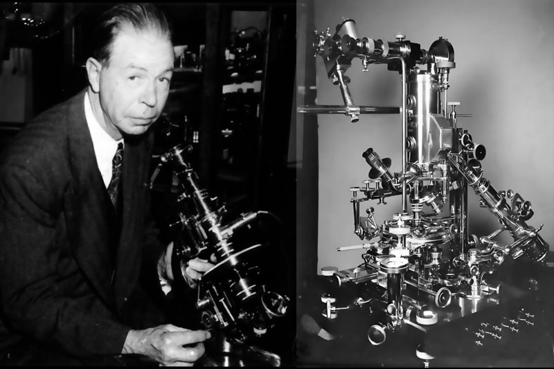
Dr. Rife aggressively pursued and achieved what had not been done in the field of ultra-microscopy. His mind had turned over the method which he had conceived so many years before. The dream which Dr. Rife originally received was now in view. Looking for more light. He decided to try filling the entire objective with cylindrically cut quartz prisms. There would be no difference in refractive index from start to finish along the optical path. Quartz prisms would “open out” each ray convergence, maintaining strictly parallel ray cadence. An increased ray content being thus returned to the ocular, the image would be brilliant in appearance and of high resolution.
This configuration of quartz prisms caused the rays to “zig-zag” in 22 light bends. The internal optical path was now entirely composed of22 quartz blocks, fitted snugly to lenses. It was as if the entire device were one solid crystal of diverse surfaces. Now, specimen emergent light would launch out in parallel paths through quartz prisms, being magnified only when they reached each quartz lens. This optical tracking method would insure the brilliance of the emergent image.
A second optical innovation was added to this brilliant configuration. Dr. Rife decided to use a phenomenon by which strong specimen-entrant light stimulates internal fluorescence in the specimen. Pumping the specimen with brilliant ultraviolet-rich light would shift the divergence point into the very heart of the specimen rather than beneath, forcing the specimen to radiate its own brilliant ultraviolet rays.
Here was a true, vanishingly small radiant source with which to illuminate the specimens: they themselves would become the radiant source! This concept was truly sublime, since the very infinitesimal particles themselves were now made to radiate brilliant and divergent rays. This scheme was truly original from the very start. Dr. Rife then designed a system by which selected portions of the ultraviolet spectrum could be split and directed into the specimen using a polarizer. Turning this component of the system would allow each specimen to brightly fluoresce in its own absorption spectrum, the infinitesimal specks radiating their own maximum brilliance.
Theoretically, it was possible to magnify these brilliant specular rays to any degree. But a secondary monochromatic ultraviolet ray would perform an unheard wonder. When combined with the brilliant internal fluorescence of the specimen, this secondary ultraviolet addition would heterodyne the light. This meant that light pitches from the specimen would be raised far above its original values. At such shorter wavelengths, the resolving power of this device would be incredible.
An additional monochromatic deep ultraviolet beam was mixed with the fluorescent radiance of specimens, producing an astounding visual sharpness of otherwise invisible objects. The illumination scheme and the tubefilled cluster of quartz prisms (designed to maintain the specimen emergent rays in absolute parallels) were now brought together. Dr. Rife claimed that these parallel lines were within one wavelength of accuracy, an astounding claim.
He soon created a small ultra-microscope whose fundamental mode of operation violated the supposed laws of optics. This design outperformed all previous ultramicroscopes. So astonishing was this feat that the Franklin Institute, in rare form, published a long and detailed series of articles concerning the developments of Dr. Rife. They were also given several of these units for preservation, where they remain to this day.
This microscope was different, totally different. This microscope revealed not just viruses in their dormancy. This microscopes could see viruses in their active stages with magnified clarity. Dr. Rife’s Prismatic Microscope surpassed the theoretical limits which were possible for optical microscopy in 1930, giving unheard resolutions of 17,000 diameters; three times the resolution developed by Dr. Lucas.
The first Prismatic Microscope was a horizontal optical bench assembly, mounted on a massive pier. Fitted with the finest photographic instruments, Dr. Rife took breathtaking photographs at unheard magnifications. The resolution was so staggering that research institutes rushed to watch Dr. Rife’s demonstrations.
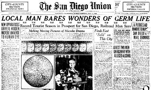 His accomplishments were extolled by the entire medical establishment on both sides of the Atlantic. An incredible amount of professional research publications devoted lengthy articles to his achievements. His findings were duplicated and reported by leading medical institutes whose names are well known. Therefore our general lack of knowledge concerning his life story is equally conspicuous.
Dr. Rife, humble enough to have worked as a chauffeur, had been raised from obscurity to fame …from shadows to light. The man’s genius was only equalled by the upstanding character by which he was loved. The Timkin family adored him. The Rife laboratory was completely equipped with the very finest apparatus money could buy. Dr. Rife methodically designed new research ventures. Incredible new biological discoveries followed him in every direction. Now, with this “ultra vision”, he was able to peer with his colleagues into unheard dimensions. These discoveries quite often challenged accepted biological and medical notions.
Dr. Rife had in mind the creation of an Institute, in which he could train younger specialists in the operation of these wonderful ultra-microscopes. Mass production of the devices would be insured. They would become fixtures in every professional laboratory. Money was not the aim of this research. Monies were already secured. Dr. Rife had a singular goal, and demonstrated the passion associated with his quest.
He developed seven different models of this initial projection-type prismatic microscope in quick succession. The horizontal projection format was converted to a more compact vertical orientation, best serving the needs of pathologists and biologists in practical laboratory settings. Several of these wonderful Prismatic models may be seen in the various archival films and photographs taken in Dr. Rife’s laboratories.
If the Rife Prismatic Microscopes outperformed every standard laboratory microscope, being able to discern and photograph virus particles in their active state, the Universal Microscope outdid all the former records. In 1933 the creation of the Universal Microscope afforded resolutions in an astounding excess of 31,000 diameters, with magnifications in excess of 60,000 diameters.
Using technically precise photographic enlargement techniques, he was able to provide 300,000 diameter magnifications. His calculations indicated that a ultra-optical projection microscope giving clarified magnifications of 250,000 diameters would be possible. After photographic enlargement, there would be no limit to the optical viewing power unleashed for researchers.
The Rule of Abbe was mentioned as a failed byword in Dr. Rife’s laboratory. He had succeeded in breaking the “vision barrier”. There are those whose familiarity with optics and attainable optical precisions state that the claimed magnification effects cannot be obtained with ordinary principles of light. Beyond simple optical parameters, other light energies become the more active in such devices. The focussing process is radionic in effect, utilizing the penetrating Od luminescence. The stimulation of special retinal modes releases the anomalous perception with its reported ultra-optical magnifications. Careful examination of the Rife Ultramicroscope reveals tubes filled with quartz prisms, identical in basic use as the patented radionic analyzers of T.G. Hieronymus (Lehr).
Viruses remained absolutely invisible to the eye when cultures were searched with the then-standard Zeiss darkfield (oil-immersion) microscope. Dr. Rife’s Prismatic Microscopes were immediately obtained by Northwestern University Medical School, the Mayo Foundation, the British Laboratory of Tropical Medicine, and other equally prestigious research groups. These models produced magnification and resolution up to 18,000 diameters.
A space composed of brilliant light, where mind illuminating light merged with light in the eyes was now opened before him. Fields, all of light. The new vision would be unstoppable. No cloak of invisibility could protect the foe now. Soon everyone would see, and the armadas of death and shadow would be vanquished. The spoils of this war would flood humanity with indescribable treasure. Life and light would again be unleashed in a world where shadow and death had reigned far too long. The immense task of cataloguing viral pathogens had begun.
QUEST
With the new Prismatic Microscope models, both he and Dr. A.I. Kendall (Northwestern University Medical School) were able to observe, demonstrate, and photograph “filterable” pathogens (viruses) in 1931. Moreover, they were perhaps first to discern the transition of these bacilli from dormancy to activity over a specific period of time. Freshly made cultures were sampled at specific stages, revealing fixed periods of quiescence and activation.
An initial tissue substrate was prepared in which bacillus typhosus was cultured. After several days’ growth, samples of this lethal culture were filtered through a fine triple zero Berkefeld “W” filter. This filtration process was repeated ten times. When viewed under the best available laboratory microscopes a turbidity was seen, but there appeared no organisms whatsoever.
Under the Rife Prismatic Microscope, polarizer adjusted, the bacilli in this sample fluoresced with a bright turquoise blue coloration. Two forms were observed, taking the researchers by surprise. Long, relatively clear and non-motile bacilli were found alongside a great population of free swimming ovoids, granules of high motility. The motile granules glowed in a selffluorescent turquoise light at a magnification of 5000 diameters.
These motile forms were transferred to a second fresh substrate, and allowed to grow for days. The same filtration process was performed. When sampled randomly before the four day period, the filtered specimen revealed something remarkable. Dr. Rife and Kendall observed relatively quiescent clear containing bright turquoise ovoids at one end. The implication was enormous. Exact transition periods were thereafter determined with precision, the entire process photographed through special attachments designed by Dr. Rife. At specific intervals of activation, the clear bacilli were discharging the turquoise motile forms into the culture. These blue ovoids were the real cause of the disease. The long and clear bacilli were only hosts. Transitions back and forth (between clear host-dormancy and motile turquoise granules) were observed and reported in the professional journals. These findings were corroborated first by Dr. A. Foord, chief pathologist at Pasadena Hospital, and later confirmed and reported by Dr. E.C. Rosenow at the Mayo Foundation (1932). The Rife Prismatic Microscope was quickly earning its reputation.
Soon, other specimens were obtained and studied by the team. Active poliomyelitis cultures were studied, the virus successfully isolated, identified, and photographed in 1932 by Rife and Kendall. In these cultures the team recognized streptococcus and motile blue forms resembling typhosus. These last reports were immediately transmitted to the Mayo Foundation and duplicated by Dr. E. Rosenow. Dr. Karl Meyer (Director of the Hooper Foundation for Medical Research, University of California) came to the Rife Research Laboratories with Dr. Milbank Johnson, examining and corroborating the stated results. The impossible and anomalous became fact. Bacilli could act as virus carriers. Furthermore, poliomyelitis victims evidenced a startling degree of typhosus-like associated virus.
Frightening implications came when comparisons between the Prismatic Microscope and the Zeiss scopes were made. All of the previous studies made with Zeiss scopes returned negative results. Such reports flooded the literature. The filtrates had been maintaining their cloak of invisibility for years. Professionals, bereft of this clarified vision, were concocting numerous speculative explanations for the appearance of these disease states. The vacuum produced by lack of visible evidence was producing erroneous theories. Many highly qualified persons, in absence of the sight required to know better, steadfastly maintained that victims of certain diseases were suffering from internally developed conditions.
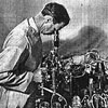
The Rife ultra-microscope was about to trigger a war on viruses. Because of the self-fluorescent “staining” method, Dr. Rife observed live specimens exclusively; a distinguishing feature of his technology. The fluorescent coloration of each pathogen was catalogued, an historic endeavor. Tuberculosis bacilli appeared emerald green, leprosy was ruby red, E.Coli were mahogany colored …each wickedly deceptive in their pretty colors. The degree of precision demonstrated in Dr. Rife’s catalogues bears the unmistakable mark of genius. We can view him at work in the archival movies.
Photographic arrays of all kinds may be seen in this footage, including the professional Scandia 35 mm movie camera with which he made stop-action films of viral incubation periods. Dr. Rife made sure to document every discovery. It was novel at the time to document every image on movie film as well as in still shots. He methodically went through every possible pathogenic specimen, photographing the deadly families. Suddenly new viral species began appearing: non-catalogued species.
The prismatic microscope was piercing into new shadows. Dr. Rife recognized unknown virus species everywhere. And then he turned his vision into the deepest shadow. He looked at the dreaded disease. To this very day the very utterance of the disease is foul. It carries the nimbus of finality. Cancer. It is an arrogant boast, a victory over cringing humanity. All who speak its name whisper in fear, afraid that it will hear and come for them. Immigrants refused to even mention the name, fearfully crossing themselves …calling it “the evil sickness”.
In the absence of fact, in the absence of vision, researchers developed contradictory theories concerning cancer and its development. These contradictory theories were eventually consolidated in the professional literature, a self-neutralizing amalgam of conjecture. Researchers were forced to examine the biochemical effects, and not the cause, of cancer. Most could not imagine what would drive cells into the bizarre and abnormal cycles common to cancer tissues. There was certainly “no visible cause”.
Dr. Rife began obtaining a wide variety of malignant tissues in 1931. The full power range of the first Prismatic Microscope was turned on these tissue samples with a vengeance. Dr. Rife was a master pathologist. His techniques can be observed in his cinematic presentations. Was he seeing correctly? What were those motile forms, glowing with a beautiful violet-red coloration? He watched them for a long while. They moved swiftly through the field of view. Clocking their motility in triple distilled water, he watched them darting across the grating. These stretching ovoids moved with startling speed.
Dr. Rife obtained yet more and diverse tumors from wider and more diverse clinical sources. An amazing 20,000 of these tissue samples were obtained and cultured. Incubating and culturing each of these required care and time. Absolute sterile conditions were maintained. He employed several groups of large high pressure steam autoclaves. No question of contamination could exist in this setting. His methods may be surveyed in the archival films which show every room of his facility. Specimens, removed from these cultures were always filtered through unused triple zero Berkefeld porcelain, mixed with triple distilled water.
Examination of each separate sample under the Prismatic Microscope revealed a consistent truth. There they were again! Always the same violet-red presence. He called it the BX virus, finding it present in every case of cancer in humans. Were these same violet-red motile forms the very cause of cancer? They were always found in every sample, a deceptive beauty. Could this be the cursed stream? Had he alone been brought to see these first? Colleagues were able to verify these findings only when using his microscopes. Both Dr. Rife and Dr. Kendall successfully demonstrated the isolation of the BX virus to more than fifty research pathologists associated with the most noteworthy institutions.
Many writers of medical theory had already postulated that there were some cancer cases which were viral in origin, but they never cited these agencies as the universal cause of cancer. The speculation, the papers, the lectures, the theories. Talk and more talk. Rife saw the universal cause of cancer. There, in full sight lay the proof positive. In case after unmistakable case, Dr. Rife found the very same agency at work. Always the same violetred motile forms. It mattered not where the tissue materials came from. There could be no mistake. There was no citing possible contaminations. Independent acquisition of tissue samples were obtained by others who then verified these findings in distant laboratories. They were using the Rife prismatic microscopes.
He succeeded in isolating the BX virus in 1931, filming the process so that posterity would hopefully learn of its enemy. He cultured this evil spawn and proceeded to demonstrate its incubation and activation periods. Transferring BX virus from culture to host, and from host to culture, all became routine. One hundred and four separate transfers were successfully made with various BX strains. Dr. Rife witnessed the appearance of another related viral cancer-causing strain, the BY virus, found to be a much larger strain of the sarcoma group. Demonstration of the infection and incubation process was subsequently affirmed by other professionals.
The same virus appeared in every case of cancer in humans. He assembled high speed movie cameras in order to clock the periods of BX virus activity. When the film ran out and was developed, he and all his colleagues could watch the deadly dance. He stepped back for a moment and surveyed the photographic evidence, flickering on the wall. Those wicked damned wriggling specks! From how many souls had they drawn away the life?
Dr. Rife now watched in horror as the malignant act was revealed before his eyes at high speed. BX virus infection required special “weakened” physiological states. Contracted as a flu-type infection, the virus incubates in host physiology for a time. When specific detrimental physiochemical states are compounded, the virus stirs into activity.
Stimulating the rapid proliferation of cell division, the BX virus forces the host body to manufacture needed nuclear material on which to further its survival. Tumors were found to be sites where BX viral colonies were rampant. Occasionally there were persons who demonstrated spontaneous remissions. These were exceedingly rare cases where antibodies actually drove off the attacking virus. Most persons could not summon this degree of response. Once the virus took control of cellular integrity, death was imminent. Shadows sweeping over humanity. There had to be a means for destroying this enemy. There had to be …a light.
SPEARS
Others, working in distant laboratories, did not claim the same success. Why had they not seen them? Because, using the over-celebrated electron microscopes, they could not see. The frightful truth concerning the BX virus was that electron microscopy could not image them at all. What had occurred in the other research labs became clear again to the man with eyes to see. The others overlooked this obvious pathogenic presence simply because their microscopes could never reveal it. This hideous specter exalted itself in what cover it could find. Unfortunately, it found cover among those who claimed to be professional seers.
Otherwise excellent researchers became completely blind when searching for the BX virus because electron microscopy was itself the blinding agent. How could so obvious a pathogen not been imaged in a technology which boasted greatest visual resolving powers? In preparing specimens for an electron micrograph, technicians “kill” the tissue specimens. The process involves placement of the specimen in a high vacuum chamber. Bombardment of the specimen with metal ions is the “staining” procedure. The thin metal film gives highly projective electrons a detailed surface upon which to impinge. The electron spray is directed into this prepared specimen and is then magnified by successive intense magnetic field coils. Images are then watched on a phosphorescent screen, or photographed directly.
Electron microscopy mishandles frail viruses. It mishandled the frail BX virus, destroying it during each preparation process. Destroyed evidence. The same ritual was repeated a hundred times with the same negative results. Unable to think clearly, few of these technicians could surmount the situation and comprehend why this virus did not appear on their viewing screens. Overconfidence in the RCA system blocked common reason. Electron microscopy does not resolve frail viruses because they are shattered and dissolved during the preparation stage.
Well then, there was the flaw. Why would no qualified person see this simple truth? Why was the light eluding those who claimed to have all of it? The technological marvel, designed to replace all competitive microscopes had brought a secure sleep on those supposed to resolve such obvious dilemmas. Medical technicians had forgotten how to think. Its newly adopted methods actually destroyed frail pathogens intended for study. Quite recently the search for the HIV virus evidenced frustration again because of these inherent limitations of electron microscopy.
The BX viruses cavorted and wriggled boldly before his eyes. But…how to destroy them? To find an immunological tool for each of these would represent an enormous task, a project which would take centuries. Humanity did not have that much time to wait. No, some other more universal means had to be developed by which this, and all pathogenic forms could be dissolved.
Protozoa and bacteria of all kinds could be destroyed by exposing them to special ultraviolet spectra. Perhaps the BX virus would succumb to such exposures. He had to know. He had the tool with which to see. So he began a long and arduous search, looking for spectra which could destroy virus cultures.
Dr. Rife discovered that deadly viruses actually thrived in the radiations of specific elements. Radium and Cobalt-60 were the notable ones. Dormant viruses became virulent in these energetic emanations. The horror filled him again. Medical practice was attempting the cure of cancer with these very radiations! There had to be some light spectra which destroyed the viral activity altogether. He search through the periodic table. Electrified argon and neon also brought intensified virulent activity from dormant viral cultures. He actually utilized argon lamps to grow virus infected tissue cultures with greater rapidity. But there had to be a spectral range which kil1ed these terrible death-agents.
No light seemed to have any effect on their crystalline structures. This is why it was possible for him to view viral activity under intense light in the first place! No light spectra of any intensity was able to destroy these quasiliving crystals.
Then he thought of crystals. How could we destroy a crystal? What do chemicals do to germs …dissolve them, take them apart…shatter them?
He had done this very thing in 1917 with protozoa and large bacteria. He knew it was possible to shatter these kinds of pathogens by the application of a sudden electrical impulse. His early attempts with small radio transmitters and simpler microscopes proved somewhat effective. He used Telefunken output tubes to produce the impulses. Operated by a small generator, this simple device projected fifty radio frequency watts to his samples.
His original inspiration applied to larger pathogens. It therefore needed no excessive frequency, short wave being sufficient. It was certainly possible to interpolate the necessarily superhigh resonant pitch needed to shatter any microbe. But viruses? How high would this pitch need to be? If not attainable,could he use some much lower harmonic of this fundamental at greater power levels? Could he find the lethal pitch for every found pathogen?
Equipment was quickly assembled. He needed a generator of extremely short duration electro-impulses. Direct current electrical “spikes” of quick duration, when applied to a gas filled discharge tube, would project electric rays toward an infected sight. The tube could not be a simple high vacuum. That would release dangerously penetrating X-Rays. X-Rays would stimulate the BX strain into increased activity. No, the projection tube required a very light gas, one whose response was almost instantaneous. The gas he desired would be one whose mass would in no way interfered with the impulses.
Hydrogen was used in special high power thyratrons: quick acting high voltage switches used in diathermy machines and (later) in radar systems. Old X-Ray tubes often failed in their operation because they became filled with hydrogen and helium mixtures. Such X-Ray tubes were generally discarded. His new projector was one such old X-Ray tube. He tested its output, adjusting the excitation circuit so as not to release even soft X-Rays. The tube glowed, a good sign. This meant that there was sufficient gas for the release of electrical rays. Dr. Rife set the polarity so that the tube would pulsate in electropositive spikes of specific duration.
Power was ready. Pathogens cavorted boldly in view. Poised at the Prismatic Microscope, he fired the X-Ray tube. Turning the tuning dial near the specimen, he would know the lethal pitch by watching the pathogens. When these “exploded” he would mark the setting. If this method worked, then he could methodically correlate each lethal pitch with its pathogen. Soon, a catalogue of lethal pitches would be amassed. With this Dr. Rife could wage victorious wars against every disease in existence.
Dr. Rife swept through the diathermy range, which he calculated should vibrate these viruses to pieces. Empirical evidence always contradicts the theoretical. Quite below the calculated extreme frequencies, the BX virus suddenly dissolved. He switched off the transmitter and sat there quite amazed. The scene in the microscope was unreal. Not a fraction of a second at the lethal pitch and the specimen was reduced to a glomular mass. The viruses were stuck together in shattered fragments! He had successfully “devitalized” them.
Fine tuned lethal frequencies now filled his catalogue. With great precision Dr. Rife determined every lethal pitch as planned. Armaments of light against legions of shadow. Analysis of the electropositive impulse showed that its radiance was penetrating, intense, and unidirectional…more like invisible light rays of pure electric force. What then was this strange lightlike power? Experiment proved that virus cultures were absolutely incapacitated, congealed, and destroyed by the electropositive impulse. The power of an extreme form of light? Had such light ever been seen before?
This energy had been accidentally generated in 1872 by Thomson and Houston. Not waves, but rays. Electrical rays. A forgotten phenomenon. Unidirectional electric impulses of great power radiated electric rays, not waves. These rays penetrated all kinds of matter, whether stone and steel alike. The resultant sparks could be drawn from every insulated metal object in the large building in which the experiment was being performed. Not radiowaves, but electric rays.
Later in that century, Nikola Tesla accidentally observed the same electric ray production. He studied the phenomenon exclusively, developing impulse generators and electric ray projectors. When speaking of electric rays which evidenced a light-like nature he referred to this phenomenon. Not radiowaves, but electric rays. New light. Dr. Rife had rediscovered this phenomenon. Tesla spoke of his own “millimeter rays”, mentioning their “bacteriocidal” value. This same phenomenon had vindicated Tesla’s words. Therapeutic properties were demonstrated when precisely controlled.
FORTRESS
Whereas the destruction of virus cultures on a quartz slide was easily accomplished, the destruction of pathogen cultures in human hosts was not. Rays had to penetrate through skin, musculature, and bone; a considerable resistance through which to travel. Rays might lose their original accurate pitch in this transit, destroying the intended action altogether.
Fortuitous and strange, the pathogens were found to be some two thousand times weaker than body cells. This meant that pathogens could be destroyed by the radiant impulse method without harming the patient. How sublime. Pathologists had treated micro organisms as chemical systems for a century, working overtime in order to find each specific chemical dissolving agent. This method treated all germs as mechanical systems, dissolving them with vibrations.
He himself had been exposed to the instantaneous blast without harm. When adjusting the rates to annihilate ordinary viral infections, he noticed that he became drowsy and tired for a few hours. Determining the cause of this as the resultant toxin release after infective agents were coagulated, he recognized the need for a de-toxifying agent. Physiology had to be prepared for the curative impulse. Exposure would release large amounts of toxic pathogen fragments into the bloodstream all at once. The ray cure had to be metered in doses. Body tissues had to flooded with special fluid electrolytes to aid the enhanced and rapid elimination of these toxins.
To stimulate deepest shattering action, the patient had to be bathed in a “carrier field”: an electrical body permeation in which the impulse light rays could penetrate into and through every body cavity. Superficial exposures would not completely cure the patient. This light ray energy had to permeate the body completely. Dr. Rife conceived of method whereby patients could be enveloped in a harmless body-permeating electric field of acoustic frequency, while the intense electro-impulses of short duration would be simultaneously projected. In this manner, efficacious electro-radiant impulses could shatter specific pathogens throughout the infected body with no harm to the patient
Dr. Rife utilized two banks of oscillators with which to generate his primary and secondary impulse fields. Acoustic generators supplied the primary field of “immersion”. A diathermy machine was coupled to a powerful transmitting amplifier to provide the shattering impulse. Two radiant energies were thus employed to destroy pathogens in vivo. Dr. Rife’s catalogue of lethal rates always gives a pair of lethal frequencies per pathogen.
Dr. Rife discovered that virus cultures were not safe from the radiant impulses from the special ray tube. Fixed to the lethal pitch of a single pathogen, the rays were unerring in their message. Selectivity was the hallmark of the Rife curative method. Several pathogens could be assembled adjacent to one another. Choosing the lethal pitch for one of these, the others would remain unharmed. The target, however, was utterly destroyed.
Dr. Rife tested the lethal effective distance of his rays, determining the safe placement of patients from the radiant source. Pathogen cultures did not seem “safe” anywhere near the device at all. Arranging the tube at one end of his laboratory, Dr. Rife brought cultures out to increasing distances from the radiant tube. In a final amazing experiment, he took cultures away from the laboratory in sealed containers. It was found that radiant tube emanations operated effectively on viral cultures up to an eight mile distance! Metal cabinets did not protect viral cultures from the deadly ray effects either, being ray conductive. Even when locked in aluminum cabinets, the entuned light-like rays destroyed their pathogens wherever found.
This represented a major medical discovery of greatest value to all humanity. This principle actually made possible curative broadcasts. Entire populations could be electrically “vaccinated” from single monitored sites. The world potential of this system was staggering. Now the outbreak of epidemics could be controlled without the time-consuming need for individual inoculations. The radiant lethal message would eradicate specific pathogens in several simple broadcasts. The constant monitoring of socially prolific germ populations could be maintained by continual public health “broadcasts”.
CONQUEST
He ran his entire staff through varied frequency exposures. Infections of all kinds each dissolved before The Ray. Dr. Rife was able to isolate the pathogens of infection and destroy them with the mere turn of a dial. The specificity of the ray tube device was so precise that singular germ strains could be individually mass-targeted. Cured by the flick of a switch!
Firing the tube in the lab provided a continual source of inoculation. After a time, so little toxicity was present in staff members’ bodies that the drowsy effects were never again encountered. They did not contract any illnesses. Not even colds.
After a time, Dr. Rife rarely used gloves when handling the viral specimens. Furthermore, neither he nor his technicians ever contracted any of the diseases which were handled. The Ray tube “inoculated” them all against every disease. He reported these findings to the community, while himself remaining the designer and developer of the system.
Dr. Rife, a research pathologist, never used these devices in medical practice. Other physicians desired the units for their own purposes, recognizing the potential for curing human suffering. Dr. Lee De Forest supervised the design and assembly of many oscillator components for the Rife System. WD. Coolidge himself (General Electric) willingly sent Dr. Rife hundreds of X-Ray tubes which were altered with a mixture of hydrogen and helium by Rife and his technicians. These improved tubes were tested so that they would project only the desired electro-impulse rays. These noteworthy references best recommended the Rife Ray tube System to medical practitioners of the day.
Hearing of these wonders, numerous physicians began requesting that smaller, more portable units be designed. Soon, Rife Ray tube devices were being assembled and given to physicians for limited use in their own practice. When properly operated, these devices returned successful reports, effecting complete eradications of infections and cures of various conditions. There were never any adverse reports concerning the Rife Ray tube Instruments. Neither could there be. Rated at such safe peak performance levels, no harm could possibly come from the portable devices.
The careful and reasonable monitoring of patient progress, the Rife frequency devices were bringing about a therapy revolution. Strep throat could be cured in an instantaneous exposure, seated in a physician’s office. A specially designed gargling solution was given to remove the resultant toxicity from the site.
In 1934 Dr. Milbank Johnson, Chief Medical Director of Pacific Mutual Life Insurance Company, established a therapy center for cancer treatment in Scripps Castle, San Diego. A staff was brought together from specific institutions including Dr. G. Dock (Professor of Medicine, Tulane University), Dr. C. Fischer (Children’s Hospital, N.Y.), Dr. W. Morrison (Chief Surgeon, Santa Fe Railway), Dr. R. Lounsberry, Dr. E. Copp, Dr. T. Burger, Dr. J. Heitger, Dr. O.c. Grunner (Archibald Cancer Research Committee, McGill University), Dr. E.C. Rosenow (Mayo Clinic). Dr. Rife functioned as a general consultant in matters of system therapy.
Using a Rife Ray tube system, the team received cancer and tuberculosis patients. Fifteen cancer patients, each pronounced hopeless by medical experts, arrived at the clinic. Each evidenced progressive states of the disease. A few patients were ambulatory. Treatments with the Rife Ray tube method were routinely applied. The dream was becoming real. Humanity was at last receiving its help.
Recognizing the critical condition of their patients, it was decided that exposure time would be raised to three minutes duration. It was discovered that exposures could not be repeated daily without necessary long rest periods. These critically ill patients could not withstand the extreme resultant toxicity released into the system as BX viruses were shattered. Emotional depression often resulted until the ray-dose was safely assessed. The team conferred hourly to assess the progress of each patient. Excessive exposure to the rays could result in severe lymphatic infections and blood poisoning. Therefore three minute treatments were repeated every third day, the rest periods necessary for blood detoxification.
Soon, the ray had done its work on the once-terminal victims. Constant blood and tissue samples revealed no BX viral presence in these now fortunate individuals. In sixty days’ treatment time, and after examination by several physicians, each was released as cured.
Though under continual surveillance, no relapses occurred. The treatment was revolutionary. The results, thrilling and complete. Moreover, they were confirmed by a special medical research committee of the University of Southern California. Three more clinics were opened with Dr. Johnson as General Medical Supervisor. Other participating physicians included Dr. James Couche, Dr. Arthur Yale, Dr. R. Haimer, Dr. R. Stafford with a mounting number of participating physicians. Clinics were operated between 1934 and 1938 having such a number of cures that it is difficult to list them all without simply reprinting the Rife files. Each of these cases were sent out and corroborated by other (non-participating) physicians.
In 1939 Dr. Rife was formally invited to address the Royal Society of Medicine, which had recently corroborated his findings. He was requested to bring all possible films, slides, and apparatus with great enthusiasm. Dr. R. Seidel reported these findings and formally announced the Rife Ray tube System therapy for cancer in the Journal of the Franklin Institute. (Vol. 237 no.2 February 1944).
The formation of the Ray Beam Tube Corporation was announced, through which several models would become available to the medical world within a short time. Highly skilled hospital staff members and leading physicians were very receptive to the proliferation of this therapy. Here was a new means for controlling and eradicating any kind of disease by the press of a switch. This therapy would inadvertently challenge pharmacological methods, raising human standards to a new and lofty height. The dream seemed ready to materialize.
INQUISITION
Rife found both himself and his staff members under a strange series of attacks by unknown agencies. During this time, and under very mysterious circumstances, Dr. Johnson died in a hospital bed. Brought there for completely minor reasons, he was found in bed. The local chapter of the Medical Association proceeded to bring Dr. Rife to the San Diego Supreme Court, but lost their case (1939). Dr. Rife could not be charged with malpractice, being a research pathologist and designer of medical instruments.
This repugnant offense unmasked the heinous resentment behind which many powerful individuals had previously been camouflaged. The court action itself caught Dr. Rife quite unaware. A visionary, his entire life had been dedicated to humanity. Alleviating human suffering was his life theme. Here now was strong evidence that factions within the Medical Establishment were actually mobilizing against proven therapeutic methods. Cancer itself and other equivalent maladies were being cured. Why then the assault?
Growing opposition from deeper factions of the Medical Association brought pressure on Rife Treatment clinic staff members. Threats and other unprofessional pressure tactics forced members to leave the team in quick succession. In campaigns clearly waged to malign Rife and his findings, the Medical Association assailed remaining participants in the clinics until Dr. Rife stood alone.
Deeper than the verbal show of malignancy by other colleagues was the horrifying and insidious motivation, the implication behind the attack. Why would anyone wish to destroy so great a world-advancement? Who was betraying civilization in this critical instance? Of all betrayals and of all personnel, who in the Medical Profession would seek the eradication of such monumental discoveries? Dr. Rife’s mind reeled under the weight of these thoughts. This was not mere resistance to a new idea in a time of ignorance. Pasteur experienced that indignity. No. This was a willful, calculated resistance in a supposed enlightened time.
Horribly shocked at the entire scenario, Dr. Rife literally became unhinged in court. Trembling and weeping, he could not come to terms with the sheer hatred and vehemence exhibited by his antagonists. “Why …why are you doing this?” he repeated. The prosecution could not have produced a better effect. Seeing this weakness as the very means by which to eradicate Rife and his discoveries, they continued to attack Dr. Rife openly. Calling him continually to the witness stand, they succeeded in destroying this frail hearted man of humble greatness. In short, the prosecution forced his total collapse.
Dr. Couche was compelled to desist operating Rife Therapy clinics under threat of malpractice. The Medical Association ruled that no society member who maintained use of the Rife Ray tube System would be permitted to continue medical practice in the United States. Morris Fishbein, major AMA stockholder, treasurer, censor, editor, and controller extended his legal arm to inform each member of the Rife team of the impending legal process. All Ray tube units would be recalled, impounded, and destroyed by Federal Court order, under penalty of fines and imprisonment.
LIGHT
All participants willingly returned their Rife units except Dr. Couche and Dr. Yale. These two surgeons later stated that for twenty-two years after this action, they continued to successfully treat and cure thousands of patients with the Rife Ray tube devices which they secretly maintained. Dr. Yale published a large and concise chronological account of patients treated and cured in his practice throughout that twenty-two year period. Notwithstanding the fact that sixty percent of severe (cancer) cases brought him were medically inoperative, incurable, and hopeless, Dr. Yale confirmed that all of these persons were yet alive and living happy, full lives.
The Rife Microscopes challenged RCA and its lucrative electron microscopes. The Rife Ray tube System would eradicate the accepted lucrative pharmacological methods everywhere. Such developments did not inspire challenged corporations. Dr. Rife developed a therapeutic means which works. This is all too evident by the rage of those who assailed him.
Systematic eradications of this priority level speak of social control on a vast and hideously deep-rooted scale. Implications necessarily involve corporate trusts and governmental agencies. The notion that disease proliferation is permitted for the continuance of pharmaceutical interests is too terrible to reasonably consider. Federal Officers came to impound the entire Rife laboratory all too late. Several faithful technicians had already purloined every piece of the priceless equipment, taking laboratory components and valuable documents across the Mexican border where they yet remain. John Crane maintains the priceless surplus.
Fishbein, the editor and chief censor of the AMA saw that Rife’s name would be stricken from all previous publications, that no professional journal would dare publish anything by Rife, and that no mention would ever be made of Rife’s achievements in formal proceedings. Inescapably linked with the pharmaceutical trusts, Fishbein’s actions were all too conspicuous.
Social control has become a dominant theme since the second World War. Modifying and regulating social thought through both legal and financial steerage has brought natural discovery and true technological development to a standstill. World changing discoveries can be made but not proliferated. Cures for diseases can be proven, but not implemented.
Has the world now entered a new barbaric and vulgar time where medical wonders have become a regulated property? The historical evidence proves out these thought lines. Balancing profit against cost, it is clear that outright cures are far less profitable than exceedingly prolonged and profit-effective “treatments”. Statistical analysis of social “disease incidence” mark the yearly expected gross earnings, a profit margin of untabulated measure.
Would the honor once laid upon the development of wondrous disease cures now be shunned, the cures themselves being suppressed at will by business managers? Would compassion for suffering humanity, concern for the elevation of human living standards on a worldwide scale no longer be a major medical theme?
World Society is driven by the unmodified flow of natural scientific discovery. At the fundamental level, such discoveries are truly socio-providential. While previous epochs simply endorsed and socialized each new natural discovery, newer attitudes have suffused the world from financial “sites of infection”.
In the past, medical discoveries were never questioned or resisted. They were always looked upon as absolutes: if a medical cure for disease was found, it was taken as it truly is…a miraculous providence. Not even the most ruthless financier would dare interrupt the flow of medical discoveries in past times. This state of ethical acumen has not continually been honored.
When the records are actually examined, when the billions of research dollars have been computed and balanced against the true research effectiveness, we find a staggering disproportion. How is it that medical research of the nineteenth century, far less equipped and funded, produced definitive cures which have become medical “standards”; while contemporary medical research, best equipped and super-funded, has not produced a single cure of equal social importance in the last thirty years? Dr. Rife had the answer toward eradicating all virus potentials. Perhaps, because it was not a pharmacological one, his devices have been “legally restrained” from social proliferation.
A few moments’ calculation reveals the effective ability of research to find a chemotherapeutic vaccine against each deadly virus. The calculated time exceeds several millennia. But Rife found the only reasonable technique for destroying any virus infection at will. The answer was not a pharmacological one. Eradication of his techniques at this early stage of development would be reasonable if one were heavily invested in chemotherapies. The systematic eradication of many such (recorded) cures is revealing.
Medical authorities have stated that “no means has been found by which viruses may be destroyed”. Recent evaluations of “recaptured” Rife Ray tube units contradict this statement. Dr. Rife treated germs as mechanical systems, not chemical systems. Vibration killed pathogens by the flick of a switch. A single such device could be easily tuned to destroy all deadly pathogens. His is the only device which can destroy viruses.
UCLA Medical Laboratories, Kalbfeld Lab, Palo Alto detection Laboratory, and San Diego testing Lab all had stated that the Ray tube System is absolutely safe to use. The FDA went out of its way to publish and maintain Federally directed rulings on the Rife Ray tube System, refusing to make further statements concerning its historically proven effectiveness in thousands of cured cancer cases.
A great gathering of esteemed colleagues of the medical and research professions came to honor and support Dr. Rife after the entire court affair. Friends who were too frightened to stand and fight at his side were now smiling, drinks elevated. But the man who was asked to stand and receive honor saw through the charades.
The seer saw the thick shadows which enveloped the professionals and other dinner guests. Armadas of pathogens were drumming their war drums again. Soon on the march, they would devastate humanity once again. It seemed that not one of the esteemed guests cared. The Rife Ray tube Therapy was the only time in history that viruses could be selectively and dynamically destroyed. No chemotherapeutic agencies were ever required in the process. The mere closing of a switch could achieve these undreamed wonders.
Dr. Rife had developed and implemented what no contemporary medical research group has ever conceived. And, by the end of World War II, was prevented from ever doing so again on American ground. The cheers and accolades rang on, while standing ovations lasted for more than fifteen minutes. The now frail and ghostlike discoverer looked away. Far off and away. Searching through the shadows, searching in his own darkness …for new light.

Related posts:

Posted in Science For The New Agewith 1 comment.
John Crane has since passed and just before he did the FDA put all of his equipment “under arrest” and hauled everything he had concerning Rife away. A friend of mine, Ben, personally witnessed this.