Atomic Radiation And Its Effects On Living Tissue ~ Part II
 by Jack Schubert, Ph.D. And Ralph E. Lapp, Ph.D.
The late effects of radiation are often produced in cooperation with disturbances in body function in general. One of these disturbances in particular—hormonal imbalance—can in itself aid and abet the cancer-producing effects of radiation or even delay for years the appearance of the cancer. Consequently in evaluation of the changes wrought by radiation in the body we must also take into account the complex inter-relationship existing between the various organs and the hormones released by the different endocrine glands. For example, irradiation of the ovaries in mice almost always leads to a large increase in ovarian tumors. However, the increased tumor incidence is not a simple cause and effect. What happens is this: radiation of the ovaries in mice with as little as 30 r depresses hormone production and stimulates the distant pituitary gland to put out excessive amounts of gonadal-stimulating hormones (GSH). The GSH act on the already disturbed ovary, with the end result that an enormous increase in ovarian tumors is observed. These can be prevented by timely administration of ovarian hormones.
Since hormones influence the regeneration and growth of almost all the cells in the body, it is reasonable to assume that many of the late changes and cancers developed in the body after radiation are related to the impairment of the endocrine glands such as the pituitary, thyroid, gonads, and adrenals, either as a result of the radiation directly on the endocrine organ or, indirectly, because of damage to a distant organ which then brings forth a response from the endocrines. Clinically one observes changes in people suffering from radiation-induced lesions which are suggestive of hormone effects. For example, a girl who worked on luminous watch dials and suffered from radium poisoning had no symptoms until she became pregnant. After the delivery of her child the symptoms (pains in the bones) subsided. She eventually developed a fatal bone cancer.
Another suggestive hormonal effect is found in the generally greater susceptibility of children to the harmful effects of radiation. Thus, as mentioned earlier, as little as 200 r of radiation to the thyroids of children has produced thyroid cancer, while none has as yet been observed in adults who received even 2000 r or more to the thyroid. However, until repeated observations of large numbers of adults with irradiated thyroids extending over at least a twenty-year period are made, no unambiguous conclusions can be reached.
One practical consideration arising from the production of cancer from irradiation is the very serious question as to whether radiation should ever be used for the treatment of benign (non-cancerous) conditions. With a very few exceptions the answer is a resounding “No!” in view of the fact that most radiation treatments of benign conditions involve radiation doses of hundreds of roentgens, and sometimes thousands. This conclusion is supported by the evidence of thousands of injured victims. One important medical report on this subject is worth citing here. In the medical journal Cancer, in 1948, Dr. W. G. Cahan and associates at the Memorial Hospital in New York reviewed their experiences with the use of x-rays in the treatment of benign bone lesions over a period of more than twenty years.
“At Memorial Hospital we have steadily inclined toward the belief that roentgen-ray or radium treatment is inadvisable for benign tumors of bone and for most associated bone conditions. . .”
After reviewing cases of eleven people who developed bone cancer many years after the irradiation of bone, they concluded that bone cancer does develop in normal bone after irradiation and they pointed out:
“A latent period of from five to more than twenty years following exposure is required for development. It is seldom seen after moderate doses and probably requires a tissue dose of 3000 r. There is little uniformity in the dosage, however; sarcoma [bone cancer] ensued in case 6 after a tumor dose of 1550 r.
“The tardy development of bone sarcoma after irradiation is another reason for believing that roentgen-ray therapy is not ordinarily to be recommended for non-malignant tumors of bone.
“We believe that more cases of this sort will be reported as the condition becomes more widely recognized.”
The effects of radiation on man involve such a complicated series of changes that in the final analysis we must depend on observations made from actual experience and experiment. The information on the effects of radiation on man and his individual organs comes from four main sources: (1) patients who have received irradiation for the treatment of cancer; (2) persons who have been damaged by radiation during the course of their occupation (these include the radium-dial workers, who have been a source of valuable information on the late effects of radioactive isotopes deposited in the body; radiologists and other doctors who have received continuous over-exposures from many years of working with x-rays, radium, and fluoroscopes; and atomic-energy workers who received accidental exposures); and (3) the survivors of the atomic bombings of Hiroshima and Nagasaki: and (4) controlled experiments on animals and other biological subjects, including one-celled animals, flies, rats, chickens, frogs, and monkeys. We must emphasize that all the injuries produced in man by radiation can be and have been produced at will in animals. To say, however, that animal experiments will provide all data applicable to human beings is not valid. For one thing, the longer life span of man means that radiation effects will be found that do not become manifest in animals of shorter life span. Furthermore, if we concern ourselves with such elusive changes as those involved in intelligence, it is obvious that animal experiments are of little value.
Most radiation exposures involve only a part of the body. For this reason we must survey the effects of radiation on individual organs, as well as the results of whole-body radiation. The harmful effects of radiation can be divided into those which develop within a few weeks after exposure and those developing months or years later. The severity of the radiation injury is the net result of several interrelated factors: the type and dose of radiation received, the duration of the exposure, the size of the area that has been irradiated, and the age of the person exposed.
Whole-body irradiation. How much radiation delivered in one single short dose to the whole body of 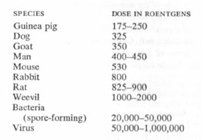 man will cause death within a month? From the Japanese victims, peacetime radiation over-exposures to individuals, and animal experiments, it can be estimated that between 400 r to 500 r would kill half of the individuals exposed. This assumes that the organs receive exposure to radiation. In peacetime such exposures would be rare except in certain therapeutic procedures involving malignancies such as leukemia. Let us now follow the reactions (called the acute radiation symptoms) which would be observed in a person receiving a lethal dose of radiation. The following description is taken from a report of the British Medical Research Council:
“The first effect . . . is a sensation of nausea developing suddenly and soon followed by vomiting and sometimes by diarrhea. In some people, these symptoms develop within half an hour of exposure; in others, they may not appear for several hours. Usually, they disappear after two or three days. In a small proportion of cases, however, the symptoms persist; vomiting and diarrhea increase in intensity; exhaustion, fever, and perhaps delirium follow; and death may occur a week or so after exposure.
“Those who recover from the phase of sickness and diarrhea may feel fairly well, although examination of the blood will reveal a fall in the number of white cells. Between the second and fourth weeks, however, a new series of ailments, preceded by gradually increasing malaise, will appear in some of those exposed. The first sign of these developments is likely to be partial or complete loss of hair. Then, from about the third week onwards, small hemorrhages will be noticed in the skin and in the mucous membranes of the mouth, which will be associated with a tendency to bruise easily and to bleed from the gums. At the same time, ulcerations will develop in the mouth and throat, and similar ulceration occurring in the bowels will cause a renewal of the diarrhea. Soon, the patient will be gravely ill, with complete loss of appetite, loss of weight, and sustained high fever. Feeding by mouth will become impossible, and healing wounds will break down and become infected.
“At this stage, the number of red cells in the blood is below normal, and this anemia will increase progressively until the fourth or fifth week after exposure. The fall in the number of white blood cells, noted during the first two days after exposure, will have progressed during the intervening symptomless period, and will by now be reaching its full extent. The changes in the blood count seriously impair the ability to combat infection, and evidence from Nagasaki and Hiroshima shows that infections of all kinds were rife among the victims of the bomb. Many of those affected die at this stage and, in those who survive, recovery may be slow and convalescence prolonged; even when recovery appears to be established, death may occur suddenly from an infection which in a healthy person would have only trivial results.”
The radiation dose which is required to kill immediately is very much greater than the LD50 dose, for which the time period of survival is 30 days. In mice, for example, it takes a total dosage of about 100,000 r to produce death within an hour. A dose of 10,000 r produces death within a few days.
Shielding of even a small part of the body can reduce mortality very much, particularly if the organ shielded is important in production of blood cells, such as the spleen. Acute lethal radiation effects can be induced by irradiation of only parts of the body, but only by larger doses than that required when the whole body is irradiated. Very interesting experiments along this line were reported at the Geneva Atoms for Peace Conference in 1955 by G. M. Frank of the Soviet Union. In the experiments described, the minimum dose of radiation by x-rays needed to kill rats was measured when the whole body was irradiated as compared to only partial body irradiation. Here are the findings:
Minimum Lethal Dose of Radiation [X-ray to Rats]
Total body——————– 800 r
Head only——————–2000 r
Lower abdomen———–3000 r
Upper abdomen———–5000 r
Chest———————–10,000 r
Human beings and animals exposed to less than lethal whole-body radiation may appear to recover, but in the long run they die prematurely. In studies of animals, some of which had been subjected to whole-body irradiation, and some not, it seemed evident, after half the population had died, that the irradiated animals had about the same incidence of diseases as did the animals not exposed to irradiation, but on average they died sooner. The most obvious causes of death in irradiated animals, such as cancer, are more often the result of irradiation of local parts of the body or of the radiation produced in a specific organ, especially such cases as the deposit of radioactive material in the bone. It appears that whole-body exposure to irradiation, whether in a large dose or small doses, causes the symptoms associated with aging.
Observations of the effect of radiation on the life span of animals of different species lead to the estimate that 1 r of exposure shortens life expectancy about one ten-thousandth of the normal life span. In man this would be 2.5 days for every 1 r. On this basis we can expect that even the normal unavoidable background radiation of about 20 r in a lifetime shortens life about 2 months on the average. For easy remembering we can assume that irradiation to the whole body or a substantial part of the body shortens life about 1 month for every 10 r of radiation exposure. While no high accuracy can be claimed, this value is at the most perhaps two or three times too high or too low. The present permissible levels of radiation for whole-body radiation exposure of 0.3 r a week would, if the preceding figures are correct, shorten life by about 4 years. While all of this my seem academic and oversimplified, there is fair evidence that radiation does shorten human life.
Female sex organs. Since a female child contains at birth all the ova she will ever use, it is very important to note that exposure of the ovaries to radiation affects eggs which are to be fertilized in the future. Thus radiation damage is preserved by the ova and may result in defective children. Even if the children appear normal, they may carry defects in their heredity makeup (the genes) which will be manifest in later generations.
The ova and related tissues are easily damaged by irradiation. Inasmuch as the human ovary is often irradiated for tumor treatment, to induce sterility or fertility, considerable information on its radiosensitivity is available. The information has been summarized by the National Research Council’s 1956 report on the “Pathological Effects of Radiation”:
“Single doses to the ovaries of 125 to 250 r may produce amenorrhea (absence of menstruation) in 50% of women. A single dose of 170 r can produce temporary sterility for a period of 12 to 36 months. A dose of 500 r produces a permanent sterility in most women, but young women may require a larger dose [up to 1000 r or more—author’s addition]. Doses between 500 r and 624 r have produced permanent sterility in 94% of a group of women.
Male sex organs. The spermatozoa, produced by the testes, develop into their mature state from very young cells called spermatogonia. The spermatogonia are among the most radiosensitive cells in the body and even 50 r or less inhibits their development. There was a case in which a temporary drop in sperm count was caused by exposure to only 12 r. Permanent sterility for the human male requires about 500 r to 600 r, while a radiation dose of 250 r may produce sterility for one to two years. It takes at least a month before sterilization becomes effective because many of the more mature spermatozoa are relatively resistant to radiation and therefore continue to their full development. Temporary sterility is due to the depletion of spermatogonia which, being especially sensitive to radiation, are killed. Fertility returns when adequate numbers of spermatogonia have matured. However, all subsequent sperm will carry an increased load of deleterious mutations which eventually will appear in future generations.
Sterilization by irradiation is not equivalent to castration. Potency and libido are not affected. The seminal fluid is produced as usual, but no living sperm are present. When the sperm return, many are abnormal at first—two heads, no head, no tail, etc.
Blood and blood-producing organs. An ounce of blood contains about 150 billion red blood cells and about 200 million white cells. There are several varieties of white cells, and those most sensitive to radiation are the lymphocytes, which constitute about 25 per cent of all the white cells. Important functions of the blood cells are to protect the body against infection, to aid the body in the repair of damaged tissue, and to promote clotting. Consequently, radiation increases the body’s susceptibility to infections and its tendency to bleed.
A single dose of 50 r to the whole body causes the number of lymphocytes to drop by one-half in about two to three days. It takes about a week for it to return to the pre-radiation level. After higher but non-lethal doses of radiation the lymphocyte drop is precipitous, and little or no evidence of recovery may be apparent for several months. In fact it may take years before the number of white cells return to normal. Another significant observation is the fact that individuals previously exposed to radiation show a greater depression of cell numbers upon subsequent radiation exposure.
Small does of radiation below the permissible levels produce significant though small depression of the white-cell numbers. This was shown, for example, at Los Alamos in 1948 by Dr. N. P. Knowlton, Jr., in a group of ten workers who received on the average 0.2 r a week of gamma radiation for 77 weeks. But still smaller doses of irradiation will produce detectable abnormalities in the lymphocytes even when the white-cell count is unchanged, as was shown first at the University of Rochester by Dr. M. Ingraham II in 1952.
Although the very small changes in the blood-cell numbers do not seem to produce any immediate ill effects, they may be the forerunners of anemia, leukemia, and other serious and fatal blood diseases. Repeated surveys have shown that leukemia—a cancer of the white blood cells leading to uncontrolled overproduction—is far more prevalent in human beings who have received radiation over-exposure. Leukemia rates ten or more times the normal rate have been observed in the Japanese survivors of atomic bombing as a result of a single exposure to radiation, and in radiologists whose radiation exposure was about 0.5 r a week for many years. The increase in leukemia in these and other people and in children receiving irradiation for benign conditions such as enlarge thymus has also been described.
Are routine blood counts of value for the detection of low levels of radiation exposures? The National Committee on Radiation Protection of the United States does not think so. They feel that the maintenance of good radiological monitoring with film badges and the like is more important. They also have found that, inasmuch as infections, colds, and other illnesses, as well as radiation, have such far-reaching effects on the blood count, it is often difficult to separate the radiation effects from those caused by minor sicknesses. On the other hand, it is still recommended that anyone overexposed to radiation have periodic systematic blood counts made, and this applies to those who run great risks of overexposure also.
There are many organs in the body responsible for blood-cell production, removal of dead cells, blood storage, and other functions which are not yet clarified. These organs include the lymph nodes and spleen, which produce mainly the lymphocytes, whereas all other white cells and the red cells are produced in the bone marrow. The latter is very sensitive to radiation and when damaged, especially by radiations emitted by radioisotopes such as radium deposited nearby, may fail to produce new cells. The resulting red-cell deficiency is known as “aplastic anemia,” one of the causes of death of the radium-dial painters.
Excerpt from Radiation: What It Is And How It Affects You
See Part I here, Part III here.

Related posts:


Posted in Other Topicswith comments disabled.